A panoramic X-ray, also known as an OPG, is a two-dimensional (2D) dental X-ray that captures the entire mouth in a single image. Unlike traditional intraoral X-rays where the film is placed inside the mouth, the panoramic X-ray machine rotates around the patient’s head to create a wide, flat representation of the jaw structure.
Description
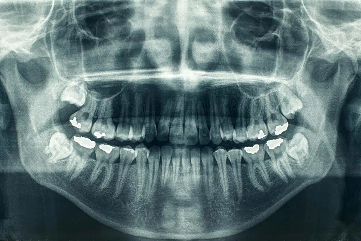
The panoramic X-ray provides a broad panoramic view of the face and teeth. It utilizes a rotating arm that holds the X-ray source on one side and the digital sensor (or film) on the opposite side. As the arm rotates around the patient’s head, it captures a continuous image of the maxillary (upper) and mandibular (lower) arches, including the temporomandibular joints (TMJ) and the nasal cavity.
Key Features
- Extraoral Technique: The imaging process happens entirely outside the mouth, making it an excellent option for patients with a strong gag reflex or those who cannot open their mouths wide.
- Comprehensive Coverage: It displays all teeth (including those that haven’t erupted yet), the jawbone, the sinuses, and the jaw joints in one frame.
- Low Radiation Dose: Compared to a full-mouth series of intraoral X-rays, a single panoramic scan typically involves less radiation exposure.
- Speed and Efficiency: The entire scanning process usually takes less than 20 seconds, providing immediate digital results for diagnosis.
Clinical Usage
Panoramic X-rays are diagnostic tools used for general screening and treatment planning rather than detecting fine details like small cavities between teeth. Common uses include:
- Impacted Teeth: Evaluating the position and angle of wisdom teeth (third molars) to determine if extraction is necessary.
- Orthodontic Assessment: Helping orthodontists see the overall alignment of teeth and the development of the jaw in children and teenagers.
- Bone Health: Detecting bone loss caused by advanced periodontal disease or identifying tumors, cysts, and jaw fractures.
- Implant Planning: Providing an initial overview of the bone height and the location of the maxillary sinuses or mandibular nerve before placing dental implants.
- TMJ Disorders: Examining the condyles of the jaw for signs of arthritis or structural abnormalities.
Comparison: Panoramic vs. Periapical X-rays
| Feature | Panoramic (OPG) | Periapical (PA) / Bitewing |
| Scope | Entire jaw and facial structure | 2–4 specific teeth |
| Detail | Lower (broad overview) | Higher (detects small cavities) |
| Placement | Outside the mouth | Inside the mouth |
| Primary Use | Surgery, Ortho, Trauma | Decay, Root canals, Bone levels |