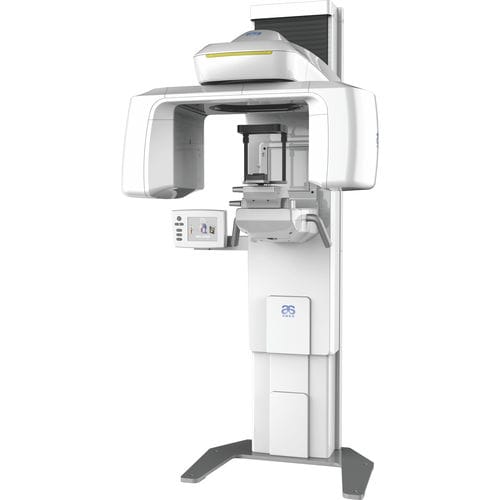
Cone Beam Computed Tomography (CBCT) is a specialized type of X-ray equipment used when regular dental or facial X-rays are not sufficient. It produces three-dimensional (3D) images of dental structures, soft tissues, nerve paths, and bone in a single scan.
Description
Unlike traditional dental X-rays which produce a flat, 2D image, a CBCT scanner rotates around the patient’s head, capturing data using a cone-shaped X-ray beam. This data is used to reconstruct a 3D representation of the patient’s anatomy. The resulting images are much more detailed than standard panoramic or intraoral X-rays.
Key Features
- 3D Reconstruction: Provides a complete spatial view of the jaw and teeth from any angle.
- Reduced Radiation: While it uses more radiation than a standard 2D X-ray, it uses significantly less radiation than a conventional medical CT scan.
- Sub-millimeter Accuracy: Offers high-resolution images (often in voxels as small as $0.075mm$ to $0.4mm$) for precise measurements.
- Rapid Scanning: Most scans take between 10 to 40 seconds, reducing the likelihood of patient movement artifacts.
- Selectable Field of View (FOV): Operators can limit the scan to a specific area (e.g., a single tooth) or capture the entire craniofacial complex.
Clinical Usage
CBCT has become an essential tool in modern dentistry for complex treatment planning:
- Dental Implantology: Accurate measurement of bone height, width, and density to determine the best implant placement and avoid vital structures like the maxillary sinus or mandibular nerve.
- Oral Surgery: Visualizing the exact position of impacted teeth (like wisdom teeth) and their relationship to surrounding nerves.
- Endodontics: Identifying complex root canal morphology, missed canals, or internal/external resorptions that are invisible on 2D films.
- Orthodontics: Assessing tooth movement, bone boundaries, and skeletal symmetry for surgical orthodontic cases.
- TMJ Assessment: Detailed evaluation of the Temporomandibular Joint for degeneration or structural abnormalities.
Comparison: 2D vs. 3D Imaging
| Feature | Panoramic X-ray (2D) | CBCT (3D) |
| Dimension | Flat, single plane | Volumetric, 360-degree |
| Distortion | Common due to magnification | Minimal to none |
| Anatomical Overlap | Structures often hide each other | Can “slice” through to see any layer |
| Nerve Localization | Estimated | Exact |