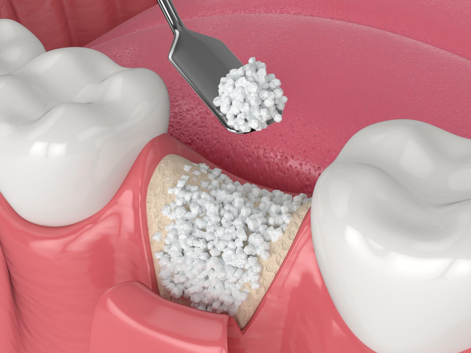
Bone Grafting Materials are substances used in oral surgery and implantology to augment or replace missing bone. They act as a scaffold, allowing the body’s natural bone-forming cells to migrate into the site and create new, healthy bone—a process known as osteoconduction.
Classification by Source
Bone grafts are primarily categorized based on where the material originates:
- Autografts: Bone harvested from the patient’s own body (e.g., chin, hip, or ramus).
- Allografts: Processed human bone from a donor bank.
- Xenografts: Bone harvested from another species, most commonly bovine (cow) or porcine (pig).
- Alloplasts: Synthetic, man-made materials such as Hydroxyapatite (HA) or Beta-Tricalcium Phosphate ($\beta$-TCP).
Description and Key Features
| Material Type | Description | Key Features |
| Autograft | The “Gold Standard.” Living bone from the patient. | Osteogenic: Contains living cells; Osteoinductive: Contains growth factors; No risk of rejection. |
| Allograft | Demineralized Freeze-Dried Bone Allograft (DFDBA). | Highly Osteoinductive; eliminates the need for a second surgical site to harvest bone. |
| Xenograft | Deproteinized sterilized bone mineral matrix. | Excellent Osteoconductive scaffold; resorbs very slowly, providing long-term volume stability. |
| Alloplast | Bioactive ceramics or glass (e.g., Calcium Phosphates). | Synthetic and Sterile; no risk of disease transmission; predictable resorption rates. |
Biological Mechanisms
To understand how these materials work, clinicians look for three biological properties:
- Osteogenesis: The material contains living bone-forming cells (only found in Autografts).
- Osteoinduction: The material contains proteins (like BMPs) that signal stem cells to turn into bone cells.
- Osteoconduction: The material acts as a physical “trellis” or “scaffold” for new bone to grow into.
Usage in Clinical Dentistry
Bone grafting is essential in several dental procedures:
1. Socket Preservation
Performed immediately after a tooth extraction. The graft material is placed into the empty socket to prevent the surrounding bone from collapsing or shrinking during healing.
2. Sinus Lift (Sinus Augmentation)
In the upper jaw, if there is insufficient bone height for an implant due to the proximity of the maxillary sinus, graft material is placed under the sinus membrane to “lift” it and create a stable base for the implant.
3. Ridge Augmentation
Used to increase the width or height of the alveolar ridge where bone has been lost due to trauma, infection, or long-term tooth loss.
4. Periodontal Regeneration
Placed around teeth affected by severe gum disease to help regrow the bone support lost to periodontitis.
Clinical Workflow
- Site Preparation: Thorough cleaning and debridement of the defect.
- Graft Placement: The material (often a mix of Allograft and Xenograft) is packed into the site.
- Membrane Coverage: A Barrier Membrane (collagen or PTFE) is usually placed over the graft to prevent fast-growing gum tissue from invading the bone space.
- Healing: Typically requires 4 to 6 months before a dental implant can be placed.